CD34
CD34(小鼠、大鼠的同源蛋白写作Cd34),是一类分化簇(Cluster of differentiation, CD)分子,在人体内由CD34基因编码,主要用来鉴定造血干细胞(HSC)等成体干细胞[5][6][7]。CD34化学本质为糖蛋白,正常情况下位于细胞膜表面,功能包括参与细胞—细胞间黏附和细胞—细胞外基质(ECM)黏附。
功能 编辑
CD34最早于20世纪80年代由两个课题组同时独立发现,最初是用来鉴定一类造血干细胞(CD34+阳性)。现在,是否表达CD34也是分离、鉴别造血干细胞及其亚群(subpopulation)的一项重要指标[8][9][10][11][12][13]。CD34为单跨膜的黏液素(Mucin)类糖蛋白,通常分布于细胞膜表面[14]。CD34功能复杂,至今CD34的功能仍然未完全阐明,已知的CD34分子功能主要是参与细胞黏附[15][16]。
分布 编辑
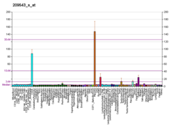
CD34蛋白主要表达于造血干细胞和造血祖细胞中,同时,血管内皮细胞、一部分间充质干细胞(MSC)也表达CD34[17]。CD34在脐带和骨髓中表达强度相对较高[18]。
参见 编辑
参考 编辑
- ^ 1.0 1.1 1.2 GRCh38: Ensembl release 89: ENSG00000174059 - Ensembl, May 2017
- ^ 2.0 2.1 2.2 GRCm38: Ensembl release 89: ENSMUSG00000016494 - Ensembl, May 2017
- ^ Human PubMed Reference:. National Center for Biotechnology Information, U.S. National Library of Medicine.
- ^ Mouse PubMed Reference:. National Center for Biotechnology Information, U.S. National Library of Medicine.
- ^ Entrez Gene: CD34 CD34 molecule. (原始内容存档于2019-09-24).
- ^ Simmons DL, Satterthwaite AB, Tenen DG, Seed B. Molecular cloning of a cDNA encoding CD34, a sialomucin of human hematopoietic stem cells. Journal of Immunology. Jan 1992, 148 (1): 267–71 [2018-02-14]. PMID 1370171. (原始内容存档于2008-06-06).
- ^ Satterthwaite AB, Burn TC, Le Beau MM, Tenen DG. Structure of the gene encoding CD34, a human hematopoietic stem cell antigen. Genomics. Apr 1992, 12 (4): 788–94. PMID 1374051. doi:10.1016/0888-7543(92)90310-O.
- ^ Civin CI, Strauss LC, Brovall C, Fackler MJ, Schwartz JF, Shaper JH. Antigenic analysis of hematopoiesis. III. A hematopoietic progenitor cell surface antigen defined by a monoclonal antibody raised against KG-1a cells. Journal of Immunology. 1984, 133 (1): 157–65. PMID 6586833.
- ^ Tindle RW. Nichols R. Chan L. Campana D. Birnie GD. A novel monoclonal antibody BI-3C5 recognises myeloblasts and non-B, non-T lymphoblasts in acute leukaemia and CGL blast crises, and react with immature cells in normal bone marrow. Leukaemia Research. 1985, 9: 1–9. doi:10.1016/0145-2126(85)90016-5.
- ^ Tindle RW. Katz F. Martin H. Watt D. Catovsky D. Janossy G. Greaves M. BI-3C5 (CD34) defines multipotential and lineage restricted progenitor cells and their leukaemic counterparts .. In 'Leucocyte typing 111: White cell differentiation antigens. Oxford University Press, 654-655. 1987.
- ^ Loken M. Shah V. Civin CI.. Characterization of myeloid antigens on human bone marrow using multicolour immunofluorescence. In: McMichael, Leucocyte Typing III:White cell differentiation antigens.Oxford University Press 630-635. 1987.
- ^ Furness SG, McNagny K. Beyond mere markers: functions for CD34 family of sialomucins in hematopoiesis. Immunologic Research. 2006, 34 (1): 13–32. PMID 16720896. doi:10.1385/IR:34:1:13.
- ^ Srivastava A, Bapat M, Ranade S, Srinivasan V, Murugan P, Manjunath S, Thamaraikannan P, Abraham S. Autologous Multiple Injections of in Vitro Expanded Autologous Bone Marrow Stem Cells For Cervical Level Spinal Cord Injury - A Case Report. Journal of Stem Cells and Regenerative Medicine. 2010 [2018-02-14]. (原始内容存档于2018-04-10).
- ^ Nielsen JS, McNagny KM. Novel functions of the CD34 family. Journal of Cell Science. Nov 2008, 121 (Pt 22): 3683–92. PMID 18987355. doi:10.1242/jcs.037507.
- ^ Drew E, Merzaban JS, Seo W, Ziltener HJ, McNagny KM. CD34 and CD43 inhibit mast cell adhesion and are required for optimal mast cell reconstitution. Immunity. Jan 2005, 22 (1): 43–57. PMID 15664158. doi:10.1016/j.immuni.2004.11.014.
- ^ Strilić B, Kucera T, Eglinger J, Hughes MR, McNagny KM, Tsukita S, Dejana E, Ferrara N, Lammert E. The molecular basis of vascular lumen formation in the developing mouse aorta. Developmental Cell. Oct 2009, 17 (4): 505–15. PMID 19853564. doi:10.1016/j.devcel.2009.08.011.
- ^ Ogawa M, Tajima F, Ito T, Sato T, Laver JH, Deguchi T. CD34 expression by murine hematopoietic stem cells. Developmental changes and kinetic alterations. Annals of the New York Academy of Sciences. Jun 2001, 938: 139–45. Bibcode:2001NYASA.938..139O. PMID 11458501. doi:10.1111/j.1749-6632.2001.tb03583.x.
- ^ Sidney LE, Branch MJ, Dunphy SE, Dua HS, Hopkinson A. Concise review: evidence for CD34 as a common marker for diverse progenitors. Stem Cells. Jun 2014, 32 (6): 1380–9. PMC 4260088 . PMID 24497003. doi:10.1002/stem.1661.


