File:Benign gastric ulcer 1.jpg
Benign_gastric_ulcer_1.jpg (400 × 408 像素,檔案大小:28 KB,MIME 類型:image/jpeg)
| 描述 |
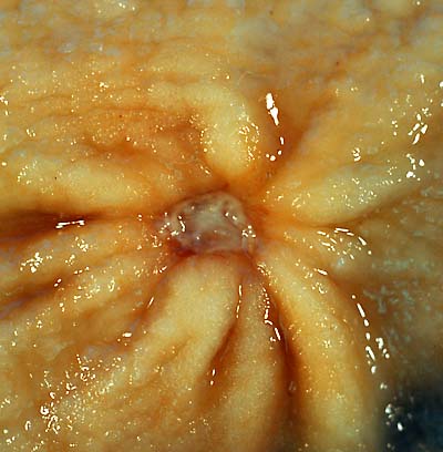
Čeština: Antrální žaludeční vřed (histologicky benigní)
English: gastric ulcer
This 1-cm benign gastric antral ulcer was discovered serendipitously in a gastrectomy specimen removed for adenocarcinoma of the fundus (not shown in the photo). The gross appearance is classic for a benign ulcer in that 1) it is relatively small, 2) the mucosa surrounding the ulcer base does not appear tumefactive, and 3) the radiating rugal folds extend nearly all the way to the margins of the base. Contrast this appearance with that of the malignant gastric ulcer included in this case collection. The criteria for grossly and endoscopically distinguishing benign ulcers from cancer are not absolute, which is why it is necessary to perform a biopsy on any non-healing gastric ulcer. Even biopsies are not 100% accurate in picking up a cancer, so negative pathology reports in such cases may provide false reassurance. The photo was taken with a Minolta X-370 with 100mm bellows lens on Kodak Elite ISO 100 film. The specimen was previously fixed overnight in formalin after being pinned out in a wax-bottomed tray. |
||
| 日期 | Posted 23 Sep 00 | ||
| 來源 | http://web2.airmail.net/uthman/specimens/index.html | ||
| 作者 | Ed Uthman, MD | ||
| 授權許可 (重用此檔案) |
|
檔案歷史
點選日期/時間以檢視該時間的檔案版本。
| 日期/時間 | 縮圖 | 尺寸 | 用戶 | 備註 | |
|---|---|---|---|---|---|
| 目前 | 2006年6月4日 (日) 22:43 | | 400 × 408(28 KB) | Patho | {{Information| |Description=gastric ulcer This 1-cm benign gastric antral ulcer was discovered serendipitously in a gastrectomy specimen removed for adenocarcinoma of the fundus (not shown in the photo). The gross appearance is classic for a benign ulcer |
檔案用途
下列2個頁面有用到此檔案:
全域檔案使用狀況
以下其他 wiki 使用了這個檔案:
- ar.wikipedia.org 的使用狀況
- ast.wikipedia.org 的使用狀況
- bn.wikipedia.org 的使用狀況
- bn.wikibooks.org 的使用狀況
- bs.wikipedia.org 的使用狀況
- cs.wikipedia.org 的使用狀況
- cv.wikipedia.org 的使用狀況
- da.wikipedia.org 的使用狀況
- de.wikipedia.org 的使用狀況
- de.wikibooks.org 的使用狀況
- en.wikipedia.org 的使用狀況
- en.wikibooks.org 的使用狀況
- es.wikipedia.org 的使用狀況
- fa.wikipedia.org 的使用狀況
- ga.wikipedia.org 的使用狀況
- he.wikipedia.org 的使用狀況
- hi.wikipedia.org 的使用狀況
- hy.wikipedia.org 的使用狀況
- hyw.wikipedia.org 的使用狀況
- it.wikipedia.org 的使用狀況
- ja.wikipedia.org 的使用狀況
- kn.wikipedia.org 的使用狀況
- la.wikipedia.org 的使用狀況
- lv.wikipedia.org 的使用狀況
- no.wikipedia.org 的使用狀況
- pa.wikipedia.org 的使用狀況
- pl.wikipedia.org 的使用狀況
- ru.wikipedia.org 的使用狀況
- ru.wikinews.org 的使用狀況
- se.wikipedia.org 的使用狀況
- sk.wikipedia.org 的使用狀況
- sv.wikipedia.org 的使用狀況
- ta.wikipedia.org 的使用狀況
- te.wikipedia.org 的使用狀況
- tr.wikipedia.org 的使用狀況
- tt.wikipedia.org 的使用狀況
- www.wikidata.org 的使用狀況