File:Electron micrograph of neuromuscular junction (cross-section).jpg
無更高解析度可提供。
Electron_micrograph_of_neuromuscular_junction_(cross-section).jpg (433 × 289 像素,檔案大小:95 KB,MIME 類型:image/jpeg)
.jpg?uselang=zh-hant)
.jpg?uselang=zh-hant)
.jpg?action=history&uselang=zh-hant)
.jpg)
摘要
| 描述 |
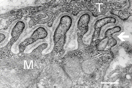
English: Electron micrograph showing a cross-section through the neuromuscular junction. T is the axon terminal, M is the muscle fiber. The arrow shows junctional folds with basal lamina. Postsynaptic densities are visible on the tips between the folds. The scale is 0.3 µm. |
| 日期 | Originally uploaded to en.wikipedia on 2006年3月10日. |
| 來源 | Synapse Web at the National Institute of Mental Health, National Institutes of Health; originally from en.wikipedia; description page is/was here. |
| 作者 | National Institute of Mental Health; originally uploaded by Nrets at en.wikipedia. |
授權條款
This image is a work of the National Institutes of Health, part of the United States Department of Health and Human Services, taken or made as part of an employee's official duties. As a work of the U.S. federal government, the image is in the public domain.
 |
||
| 此作品無已知的著作權限制,亦不受所有相關和鄰接的權利限制。 | ||
原始上傳日誌
(All user names refer to en.wikipedia)
- 2006-03-10 20:07 Nrets 433×289×8 (97758 bytes) Electron micrograph showing a cross section through the neuromuscular junction. T is the axon terminal, M is the muscle fiber. The arrow shows junctional folds with basal lamina. Postsynaptic densities are visible on the tips between the folds. Scale is 0
檔案歷史
點選日期/時間以檢視該時間的檔案版本。
| 日期/時間 | 縮圖 | 尺寸 | 使用者 | 備註 | |
|---|---|---|---|---|---|
| 目前 | 2007年3月22日 (四) 03:41 | | 433 × 289(95 KB) | Fran Rogers | {{Information |Description=Electron micrograph showing a cross section through the neuromuscular junction. T is the axon terminal, M is the muscle fiber. The arrow shows junctional folds with basal lamina. Postsynaptic densities are visible on the tips be |
檔案用途
下列頁面有用到此檔案:
全域檔案使用狀況
以下其他 wiki 使用了這個檔案:
- ar.wikipedia.org 的使用狀況
- cs.wikipedia.org 的使用狀況
- de.wikipedia.org 的使用狀況
- es.wikipedia.org 的使用狀況
- fa.wikipedia.org 的使用狀況
- gl.wikipedia.org 的使用狀況
- he.wikipedia.org 的使用狀況
- ko.wikipedia.org 的使用狀況
- pt.wikipedia.org 的使用狀況
- ru.wikipedia.org 的使用狀況
- uk.wikipedia.org 的使用狀況
.jpg)