File:Crystal structure of human CD9.pdf
此为最大尺寸。
Crystal_structure_of_human_CD9.pdf (750 × 600像素,文件大小:1 MB,MIME类型:application/pdf)
摘要
| 描述 |
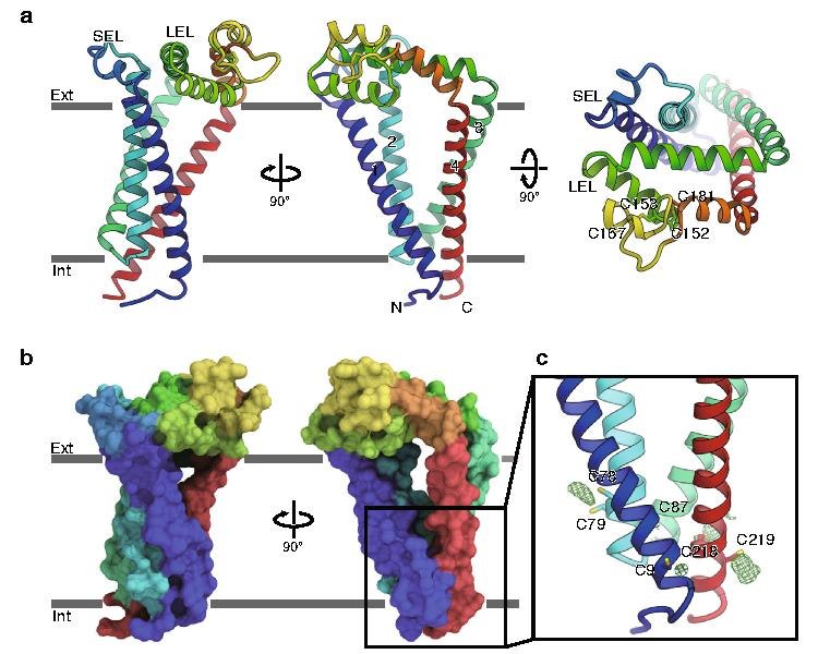
English: a Overall structure of human CD9, viewed from the membrane plane (left and middle) and from the extracellular side (right). The transmembrane helices and the two extracellular loops (SEL and LEL) are labeled. Cys152-Cys181 and Cys153-Cys167 form disulfide bonds. b Surface representation of CD9, colored according to a. c Palmitoylation of the cytoplasmic cysteine residues. Green meshes show Fo−Fc densities contoured at 2.5 σ, indicating the palmitoylation of the cysteine residues on the cytoplasmic end of the four transmembrane helices.[1] |
| 日期 | |
| 来源 | https://doi.org/10.1038/s41467-020-15459-7 |
| 作者 | Rie Umeda, Yuhkoh Satouh, Mizuki Takemoto, Yoshiko Nakada-Nakura, Kehong Liu, Takeshi Yokoyama, Mikako Shirouzu, So Iwata, Norimichi Nomura, Ken Sato, Masahito Ikawa, Tomohiro Nishizawa & Osamu Nureki |
许可协议
文件历史
点击某个日期/时间查看对应时刻的文件。
| 日期/时间 | 缩略图 | 大小 | 用户 | 备注 | |
|---|---|---|---|---|---|
| 当前 | 2020年4月4日 (六) 22:47 |  | 750 × 600(1 MB) | Rob Hurt | Uploaded a work by Rie Umeda, Yuhkoh Satouh, Mizuki Takemoto, Yoshiko Nakada-Nakura, Kehong Liu, Takeshi Yokoyama, Mikako Shirouzu, So Iwata, Norimichi Nomura, Ken Sato, Masahito Ikawa, Tomohiro Nishizawa & Osamu Nureki from https://doi.org/10.1038/s41467-020-15459-7 with UploadWizard |
文件用途
以下页面使用本文件:
全域文件用途
以下其他wiki使用此文件:
- en.wikipedia.org上的用途